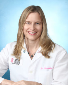
 Information about breast cancer screenings can be confusing. It seems like new recommendations are coming out all the time, and each recommendation is different from the last one. Michelle Price, MD, co-medical director of the Fortunato Breast Health Center, helps us to understand some important breast health issues.
Understanding screening guidelines
Dr. Price says, “think of it as an annual check-up. Unless there are other factors, women should begin screening mammography at age 40, and continue annual screening every year thereafter.” Many professional societies involved with the diagnosis and treatment of breast cancer also continue to recommend annual screening mammography starting at age 40, including the Society for Breast Imaging, American College of Radiology and National Comprehensive Cancer Network.
According to Dr. Price, the Fortunato Breast Health Center also does not recommend a particular age for stopping screening. “Some organizations suggest discontinuing mammography screening at age 75, but as long as you are in reasonably good health, you should continue to have your annual mammogram.”
In certain instances, women should begin mammography screening even younger than age 40. Typically, this situation would occur if a woman has a first-degree family member, such as mother or sister, who was diagnosed with breast cancer younger than 50 years of age. Having a family history of breast cancer may increase one’s risk for developing breast cancer. However, it is important to remember that 75 percent of women diagnosed with breast cancer have no family history at all. “The fact is that all women are at risk. All women can benefit from regular mammography screening and early detection. Women with additional risk factors such as dense breast tissue and family history may also benefit from complementary screening methods, such as breast ultrasound or breast MRI,” said Dr. Price.
Dense breasts – what does that mean?
Nearly half of all women age 40 and older who get mammograms are found to have dense breasts. Dense breast tissue refers to the way breast tissue looks on a mammogram. It is a normal and common finding. On a mammogram, dense breast tissue appears as a solid white area, whereas fatty tissue is more gray. Since dense breast tissue is more difficult to see through on a mammogram, it can make it more difficult to identify breast cancer. This is why the Fortunato Breast Health Center recommends that women with dense breasts have an ultrasound along with their mammogram.
A breast ultrasound can help find some breast cancers that cannot be seen on a standard digital 2-D or 3-D mammogram. Ultrasound can allow your doctor to see through the breast tissue in a way that x-rays from a mammogram cannot. Ultrasound uses painless sound waves to evaluate the tissue. These images can help your doctor identify a small breast cancer or distinguish if a mass seen on a mammogram is actually a solid lump or a fluid filled cyst. If unsure, all women should ask their doctor if they have dense breasts and if they should consider a breast ultrasound in addition to their annual mammogram.
Although supplemental screening tests are important, it is critical to remember that mammography remains the gold standard imaging test for early detection of breast cancer. Many breast cancers can be seen on a mammogram even in women who have dense breast tissue. For example, mammography is the only way to identify calcifications. It is very important that supplemental screening be done in addition to, not as a replacement for, a mammogram to rule out breast cancer.
If a woman has a screening mammogram that shows an area of concern, she is called back for a more detailed examination of the area in question. Approximately 10 percent of women screened may need this additional workup, which is called a diagnostic mammogram. Additional mammography imaging, and possibly a breast ultrasound, is then performed with the radiologist present to better evaluate the area of concern.
Sometimes an area of calcification or a breast mass is found, and needle biopsy may be recommended. We perform all types of needle biopsies at the Fortunato Breast Health Center. This is the best way to get a conclusive answer regarding the imaging finding. Nearly all patients who undergo needle biopsy report that it wasn’t as bad as they thought it would be. We take special care to provide support to our patients throughout the procedure, and many even say it was completely painless.
In the United States, the vast majority of breast needle biopsies have a benign result. Although undergoing this type of workup may be understandably stressful to patients, it’s important that any cancer be found at the earliest stage possible. Early diagnosis offers a woman her best chance at survival. “From extensive experience with our patients, we find that they are willing to endure a brief period of stress if it could be a life-saving choice,” Dr. Price said.
“There is no question that an early stage diagnosis offers the best prognosis,” notes Dr. Price. “We are well aware that mammography screening can be stressful, and that women are always busy with so many responsibilities. But please, give yourself the gift of an annual mammogram. It just might save your life.”


