

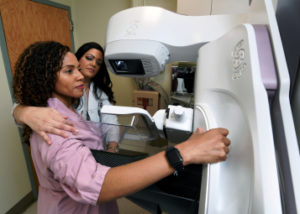
 Nearly half of all women age 40 and older who get mammograms are found to have dense breasts. Dense breast tissue refers to the way breast tissue looks on a mammogram. It’s a normal and common finding. Breast tissue is made up of milk glands, milk ducts, and supportive tissue (dense breast tissue), and fatty tissue (non-dense breast tissue). When viewed on a mammogram, women with dense breasts have more dense tissue than fatty tissue.
On a mammogram, non-dense breast tissue appears dark and transparent. Dense breast tissue appears as a solid white area on a mammogram, as do some abnormal breast changes, such as calcifications and tumors. Since dense breast tissue is more difficult to see through on a mammogram, it can make it more difficult to see and diagnose breast cancer. This is why women with dense breasts are often recommended to have an ultrasound with their mammogram.
According to a study in the Journal of the American Medical Association 3-D mammography, also known as digital tomosynthesis, has decreased some of the call-back recommendations for patients with dense breasts to have additional screening. A 3-D mammogram takes multiple pictures of the breast, which allows doctors to scroll through images of the breast tissue. This can be beneficial in women with dense breast tissue because it can reduce some of the overlapping that occurs in images of breast tissue, which can help radiologists differentiate between abnormalities and normal tissue. However, it is currently still recommended that women with dense breasts consider additional imaging tests.
Most commonly, a breast ultrasound may be recommended in addition to an annual mammogram. This test can help find some breast cancers that cannot be seen on a standard digital 2-D or 3-D mammogram. Ultrasound can allow your doctor to see through the breast tissue in a way that x-rays from a mammogram cannot. Ultrasounds use sound waves to look at the tissue and can help your doctor see a small breast cancer, or distinguish if a mass seen on a mammogram is actually a solid lump or a fluid-filled cyst.
Although supplemental screening tests are important, it is critical to remember that mammography remains the gold standard imaging test for early detection of breast cancer. Many breast cancers can be seen on a mammogram even in women who have dense breast tissue. For example, mammography is the only way to identify calcifications. It is very important that supplemental screening be done in addition to, not as a replacement for, a mammogram to rule out breast cancer.
In New York State, facilities that provide mammograms are required to notify patients if they are found to have dense breasts. If your mammogram report says that you have dense breast tissue, talk with your doctor about what this means for you. Based on your medical history and risk factors, your doctor may recommend that your personal screening plan include additional testing.
Dr. Michelle Price from the Fortunato Breast Health Center reminds, “Our goal as breast radiologists is to identify tumors as early as possible, when breast cancer is most treatable. Early detection is the best protection.”